


Treasure the little things, support your data with vivid images and reach a wider audience.
At Jungholm Laboratories, we have affordable access to state-of-the-art imaging techniques. From low-resolution high content screening to super-resolution live-cell imaging, 3D representations and more.
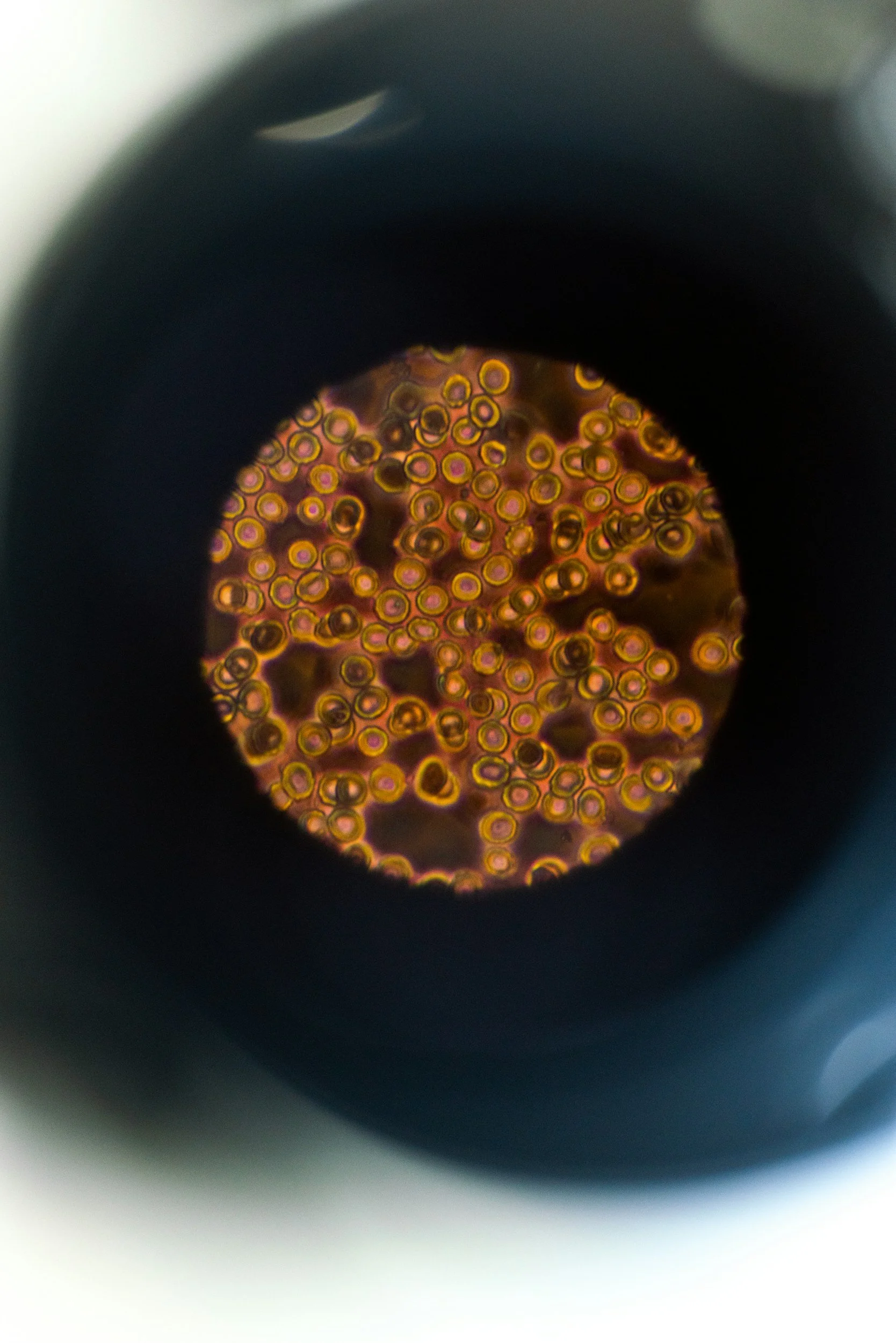
Red Blood Cells seen through the eyepiece of a microscope
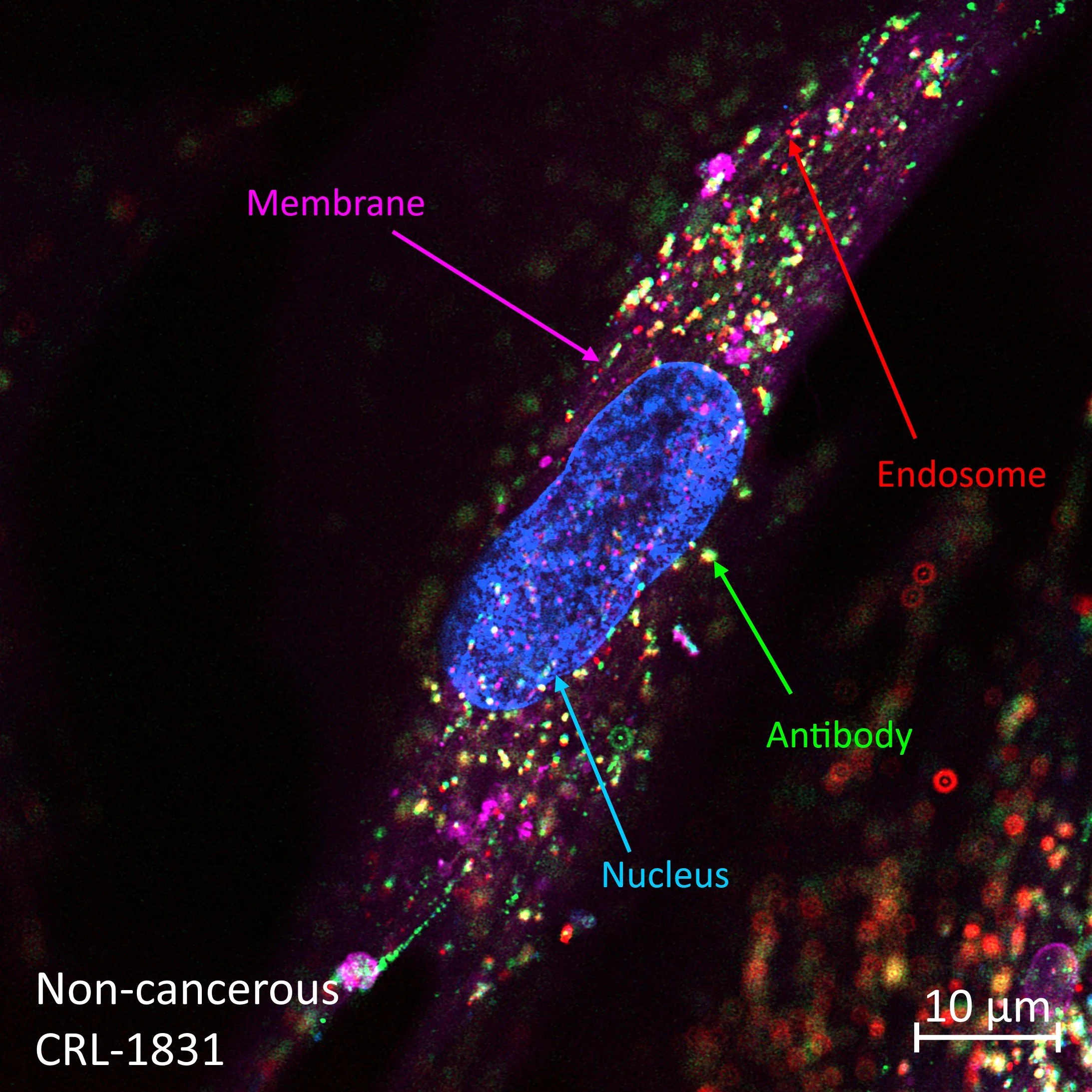
Internalization of fluorescent antibody into cancer cell, labels indicating its membrane in purple, nucleus in blue, endosome in red, and antibody in green.
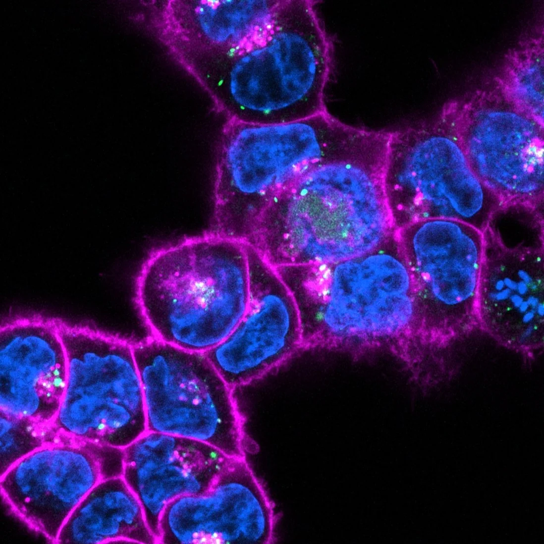
Macropinocytosis marker (green) shows distinct regions in cells responsible for the uptake of large molecules.
Communication between cells via membrane nanotubes.
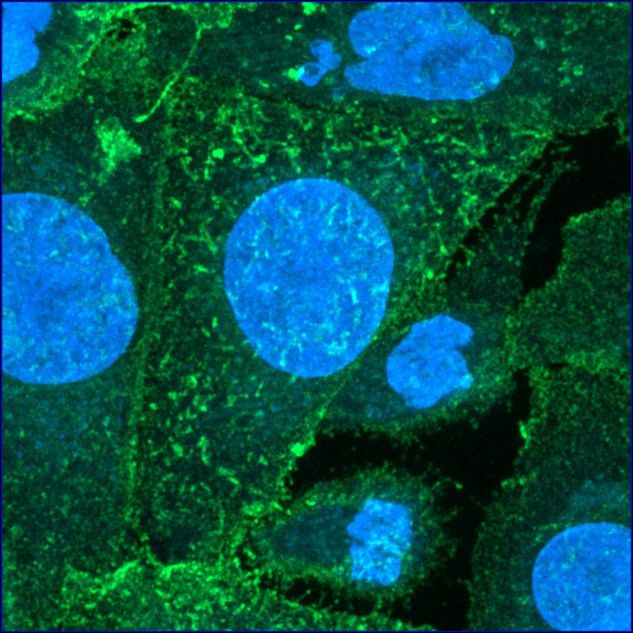
Cell line over-expressing the pain receptor TRPV1 (green).
3D rendering of a tumour organoid
Transportation of Fluorescently tagged antibody (green) in endosomes (red) within a cancer cell. Colocalization of antibody and endosome (red and green signal) is demonstrated as a histogram along a line of interest.
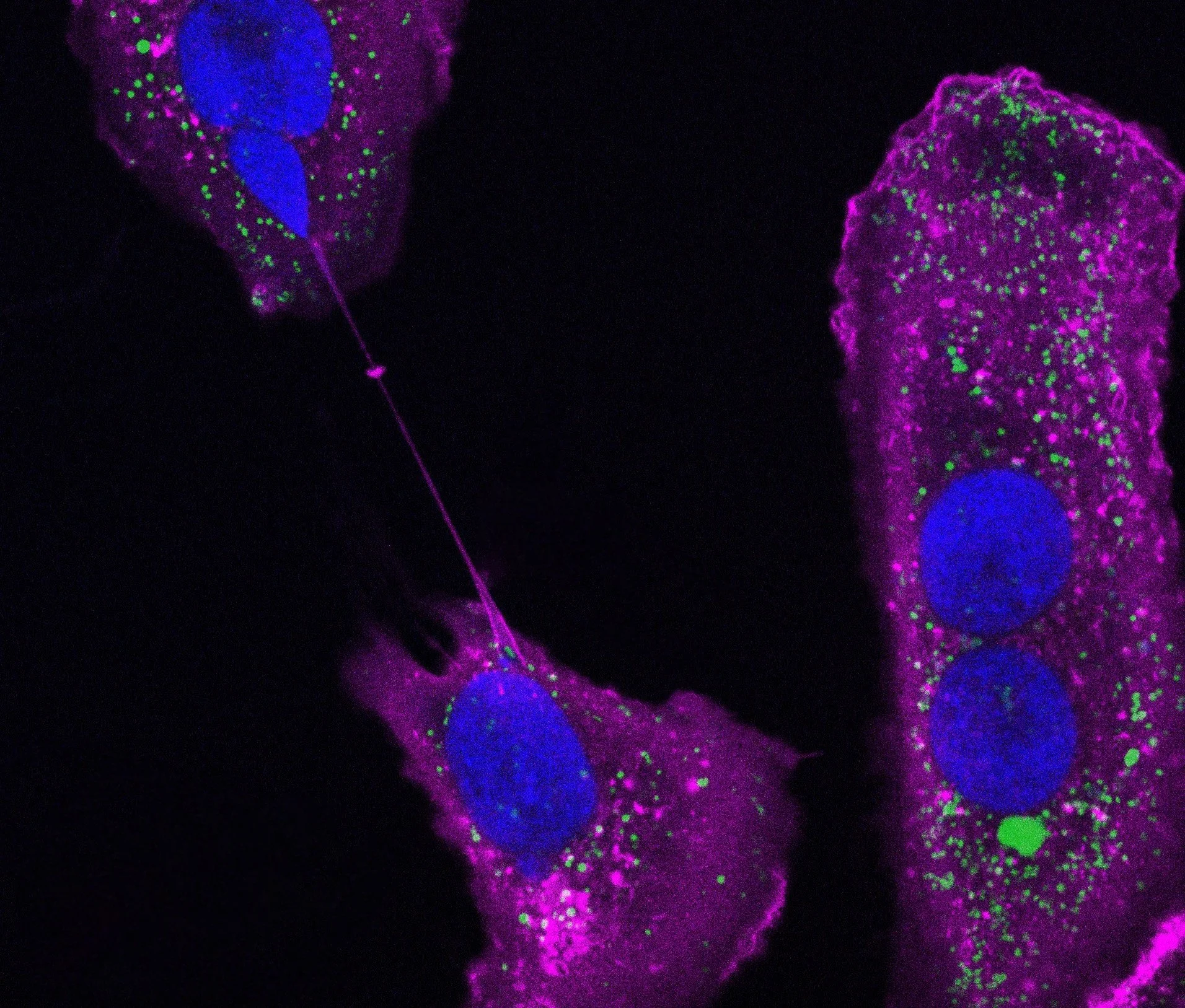
Identification of active macropinocytosis, with characteristical tent-pole formation and enveloping of the extracellular environment.
Tumour organoid, staining reveals the necrotic core.